Ridge split procedure with ridge wider and simultaneous implant placement of IS III active fixtures
- Dr. YoungKu Heo

- Mar 27, 2017
- 2 min read
Situation
A 58-year-old female patient had lost teeth 46 and 47 and was referred to the clinic for treatment.
As extremely narrow ridge was found on the mandibular right quadrant, ridge split was performed and decided to place IS-III active implant.

Pre-operative Panorama
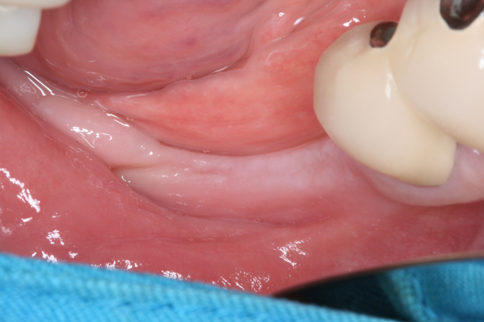
Intra-oral photograph

Flap reflection

Bone Trimmer was used to flatten and smooth out the narrow alveolar crest.
Side blades enable quick trimming of the alveolar crest.
Recommended speed when using the Bone Trimmer : 1,200rpm

Ø1.5 Initial Drill was used for initial drilling.
Recommended speed when using the Ø1.5 Initial Drill : 1,200rpm

Safe Disk was used for cutting across the narrow ridge.
Recommended speed when using the Safe Disk : 1,200rpm

Safe Disk was used for cutting the buccal side.

Safe Disk was used for cutting the buccal side.

Safe Disk Ø7.0mm, T1.0mm was used for cutting the buccal side.
✔ Ø7.0mm, T1.0mm Safe Disk is used before alveolar bone expansion.

Prevent alveolar bone fracture by cutting the lower part of the buccal area by 1mm thickness before bone expansion.

Bone Chisel was used for initial ridge expansion.
Slight expansion of the ridge through inserting the Chisel between the cortical plates.
Additionally separates the attached remaining bone

Bone Expander was used for sequential bone expansion.

Recommended speed when using the Bone Expander : 25~35rpm

CMI IS-III active (Neobiotech, Korea) 4.5*10mm fixture was placed in the area of #46.

Neo EZ-GBR Kit Ø1.5 Drill was used for insert fixing screw.

A 7mm Fixing Screw was inserted.
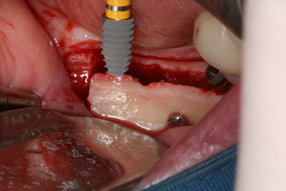
CMI IS-III active (Neobiotech, Korea) 4.5*8mm fixture was placed in the area of #47.

2 implants were placed with 15Ncm of initial stability.

Bone graft was performed.

Resorbable collagen membrane was placed over the graft.

After the surgery.

Post-operative panorama.
Click on the image to see clinical video.


Comments