Predictable Stability in Limited Vertical Bone Height: ALX-IT Short Clinical Case

Updated: Jul 13

Case Summary 🔎

Patient Information
59-year-old male
Medical History : Coronary artery disease (coronary stent placement in approximately 2012), Type 2 diabetes mellitus
Current Medication: Aspirin, antidiabetic medication(s)
Diagnosis
Treatment Plan
Placement of two ALX-IT short implants (6 mm or 5 mm) with simultaneous bone grafting
Delivery of the definitive zirconia prosthesis
Case Overview
This case presents the rehabilitation of the mandibular right posterior region (#46 and #47) with severe bone loss using ALX-IT Short implants following removal of failed implants. Due to limited vertical bone height and close proximity to the inferior alveolar canal, a short implant approach was selected to avoid extensive vertical augmentation. Following implant removal and staged guided bone regeneration, two ALX-IT Short implants were placed with simultaneous bone grafting to manage the residual peri-implant bone defects. Stable primary fixation was achieved, and the healing protocol for each implant was selected according to the individual clinical condition. Final impressions were taken 3 months after implant placement, followed by delivery of the definitive restorations. This case highlights the clinical utility of ALX-IT Short implants in a severely resorbed mandibular posterior region with limited vertical bone height
Case Presentation
1️⃣Pre-op
Preoperative panoramic radiograph demonstrating peri-implant bone loss around the implant at #46 and severe periodontal bone loss associated with Grade III mobility at #47.
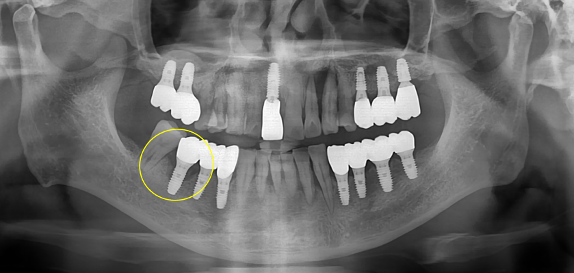
Panoramic radiograph following explantation of removal the implant at #46, with #47 temporarily retained despite severe periodontal bone loss.

Implant placement was planned 5 months after explantation at #46 and 1 month after tooth extraction at #47.

2️⃣Surgery
Intraoperative views of the #46–47 region demonstrating a severely resorbed posterior ridge, the underlying bone defect after flap elevation, and placement of drilling.

An 8-mm drill fitted with a 2-mm stopper to limit the drilling depth to 6 mm was used according to the bone quality at #46 (D333) and #47 (D002). Radiographic verification confirmed the osteotomy position and demonstrated less than 6 mm of residual bone height above the inferior alveolar canal at #47.

An ALX-IT Ø6.0X6mm, Cuff 2mm implant was placed at #46 with an insertion torque of 40 Ncm. At #47, an ALX-IT Ø6.0X5mm, Cuff 3mm implant was placed with an insertion torque of 15–20 Ncm. After confirmation of implant positioning, simultaneous bone grafting was performed to manage the peri-implant defects.

Postoperative radiographs confirming optimal implant positioning in the right mandibular posterior region, with an appropriate relationship to the inferior alveolar canal.

3️⃣Prosthesis
An intraoral scan was performed 3 months after implant placement, with IST values of 82 at #46 and 78 at #47.

An intraoral scan was performed 3 months after implant placement.

Final zirconia prosthesis delivered at 3 months after implant placement.
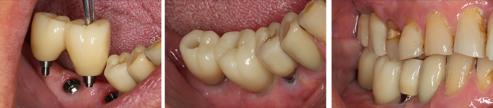
Final panoramic and periapical radiograph


Comments