Guided bone regeneration for horizontal and vertical ridge augmentation in the anterior maxilla
- Dr. Boussetta Kilani
- May 14, 2015
- 1 min read
Situation
Patient Information
- Patient: 50 year old patient
- Medical history: well controlled hypertension, non smoker
Pre-operative Observation


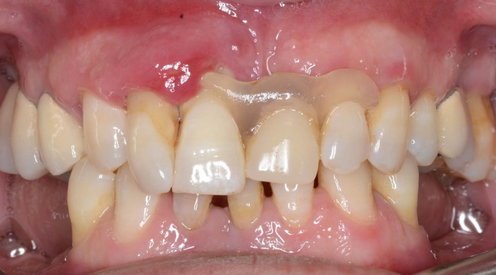

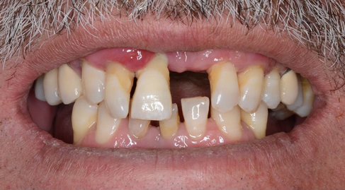


- Initial situation with a high lip position (left), Intra oral view of the area (right)
- Missing tooth on #21
- Periodontal disease
Treatment Planning
- Horizontal and vertical augmentation of the anterior maxilla with a GBR procedure
- Implant placement into the augmented ridge of site 11 and 12 in four months
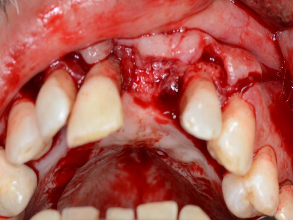
The site exposed by rising a trapezoidal flap

Frontal view of the bone defect.

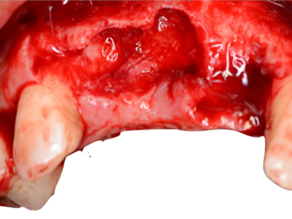
Occlusal view of the bone defect.

Perforation of the cortical plate of the bone to allow the defect volume to be populated with new vessels.

Tenting screw placement.

Occlusal view of the tenting screws.

Frontal view of the tenting screws.

The autologous bone was harvested in the cortical of mandibular area with ACM bone collector from Neobiotech, the autologous was soaked in blood and mixed with DBBM.

DBBM and autologous bone packed into the defect around the tenting screw in a slightly over contoured fashion, a resorbable membrane was used to separate the tissue compartment made up of bone from the overlying soft tissue

Tension free Primary wound closure.

Second procedure after 4 months.

Intra-operative view of the second surgical session. All the vertical and horizontal defect was resolved.

Removing of the tenting screws.

Drilling for implant placement.

Occlusal view of the implants.

End of the second stage surgery.


Comments