Fixture Removal, ALX-IT Implant Placement, and Guided Bone Regeneration


Case Summary 🔎
Patient Information
Age / Sex: 62-year-old male
Medical History: Currently taking antihypertensive medication and medication related to previous heart valve surgery.
Clinical Diagnosis: Fracture of the implant screw at site #24, requiring removal of the fractured screw.
Clinical Summary
A screw fracture occurred in the existing implant at site #24, compromising the structural integrity of the implant-supported restoration and necessitating removal of the existing fixture. The fractured implant was carefully removed, followed by placement of a new ALX-IT implant at the same site. After a 3-month healing period, clinical and radiographic evaluations were performed, and an intraoral scan was obtained for definitive prosthetic fabrication. Subsequently, a cementless implant-supported prosthesis was delivered, completing the prosthetic rehabilitation.
1️⃣Pre-op


#24 Radiographic examination revealed a fractured screw fragment retained within the implant fixture, compromising the integrity of the implant connection.
2️⃣Surgical phase

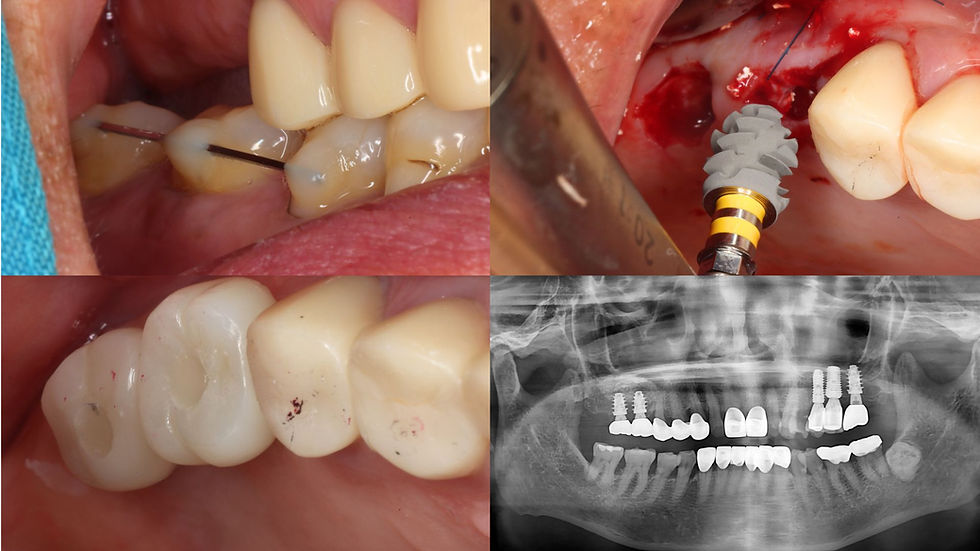
The upper portion of the fixture was severely damaged, preventing proper connection of the fixture remover.

Remove the remaining fixture using a surgical bur and an forcep.

Appearance of the removed fixture.
Surgical Report


Using VARO Guide and VARO Guide KIT


Drilling was performed sequentially from Ø2.2 mm to a final diameter of Ø3.5 mm.


The areas around the implants with insufficient residual bone were augmented using Neo-OSS B, a bovine-derived xenograft material, for guided bone regeneration (GBR).


Post-operative clinical and radiographic assessment, healing abutment placement.
3️⃣Prosthetic phase

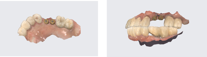


A screw-retained abutment was connected, followed by seating of the cementless prosthesis.

The cementless prosthesis was seated.


Post-prosthetic panoramic radiograph


Comments