Early Loading of an ALX-BT Implant in Soft Posterior Maxillary Bone


Patient Information
65-year-old female
Medical History: Healthy
Diagnosis: #27 healed edentulous site (Extraction 16/1/2026)
Treatment Plan
#27 ALX-BT implant placement
Undersize drilling & Sinus floor preparation for bicortical stability
Early loading of single full metal crown implant
Case Summary 🔎
This case presents the placement and early loading of an ALX-BT implant in the #27 region of a 65-year-old female patient, four months after tooth extraction.
The posterior maxilla exhibited approximately 8–9 mm of residual bone height with D442 bone quality and was classified as Dr. Heo's Sinus Classification II.
Despite the compromised bone quality, favorable primary stability was achieved with a Ø5.0 × 10 mm ALX-BT implant using a one-step undersized drilling protocol. The implant design, combined with fixation at the inferior cortical wall of the maxillary sinus, provided sufficient stability without the need for additional sinus bone grafting.
Based on the excellent primary stability, a prosthetic impression was taken 2 weeks after implant placement, followed by delivery of the final restoration at 3 weeks. At 3 weeks after loading, the implant and restoration remained clinically stable, highlighting the potential of the ALX-BT implant to achieve reliable fixation and facilitate early loading even in challenging soft-bone conditions.
Case Presentation
1️⃣Pre-op
Intraoral photograph showing the #27 edentulous area with well-preserved gingival contour and an adequate width of keratinized tissue.

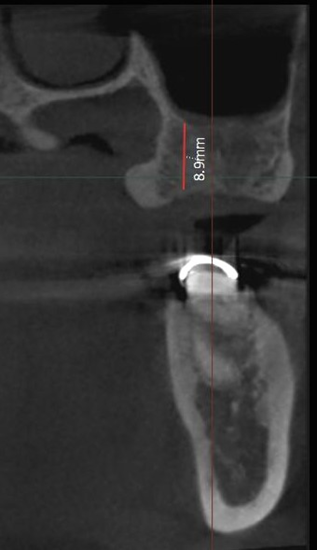
CBCT evaluation of the #27 region: residual bone height of 8–9 mm, bone width of 12 mm, and D442 bone density.

2️⃣ Surgery
Tissue punch followed by sequential drilling with Ø2.2, Ø3.0, and Ø3.5 mm drills to a depth of 10 mm from the crestal soft tissue level. A Ø3.6 mm S-Reamer was then used to prepare the osteotomy to the sinus floor at a depth of 11 mm.
A Ø5.0 × 10 mm ALX-BT implant (Ø4.5 mm neck diameter) was placed in the #27 region. According to Dr. Heo’s Sinus CMI Classification, Class II fixation was achieved through apical engagement of the inferior cortical wall of the maxillary sinus, providing favorable primary stability without the need for sinus bone grafting.
☑️Dr. Heo's Sinus Classfication
Class I CMI fixation: CMI fixation in the posterior maxilla without engaging the inferior cortical wall of the sinus (Fig 1)
Class II CMI fixation: CMI fixation in the posterior maxilla with engagement of the implant apex into the inferior cortical wall of the sinus without bone grafting into the sinus (Fig 2)
Class III CM/C fixation: CM or C fixation in the posterior maxilla with sinus elevation and bone graft through a crestal approach (Fig 3)
Class IV CM/C fixation: CM or C fixation in the posterior maxilla with sinus elevation and bone graft through a lateral approach (Fig 4)

A Ø6.0 mm healing abutment (cuff 5 mm) was connected to the #27 ALX-BT implant. Implant stability was measured using AnyCheck, with an IST value of 77 (+2).
3️⃣Prosthesis
At 2 weeks after implant placement, an IST value of 78 (+2) was recorded.
A scan body was connected to the #27 ALX-BT implant, followed by a digital impression for fabrication of a full-metal crown from a 14 mm titanium block.
At 3 weeks after implant placement, an IST value of 78 (+2) was recorded before prosthesis delivery. The final restoration was then delivered.
Intraoral photograph after final restoration delivery.
4️⃣Follow up
3-week follow-up after final restoration delivery.


Comments