The staged approach on atrophic ridge on the posterior maxilla using Neobiotech IS-II active fixture
- Dr. Seok Yong Kim

- Dec 18, 2017
- 2 min read
A 45 years old male patient, who had missing posterior teeth, presented with a partially edentulous maxilla. Implants placement on #13,#14,#15 and #16 following lateral sinus augmentation was planned…
Patient information
Male
45 yrs old
Non-contributory
NKDA
Drink occasionally, smoke 1 pack a day
PDH
Scaling and subgingival curettage PFM bridge on #23,24-26 Implant placement on #32, 36, 37, 42 Extraction on #13,14,15,16
Diagnosis & Treatment Plan
Generalized severe chronic periodontitis
Treatment plan
Scaling and subgingival curettage
Sinus augmentation using xenograft
Implant placement on #13,14,15,16

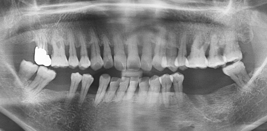
Panorama taken at the initial visit severe vertical bone resorption has been appreciated on the right posterior maxilla

Occlusal view of the surgical site before sinus augmentation

Sinus window preparation with the help of piezoelectric devices

Sinus augmentation has been performed with the use of xenograft(porcine origin)and resorbable membrane.

Single interrupted suture has been done to adapt the buccal and lingual flaps.

Panorama taken six months after sinus augmentation before implant placement.
The augmented bone can be easily appreciated.

Occlusal view of the surgical site taken just before implant surgery.

The implant placement has been done on #13,14,15,16 using Neobiotech IS-II active fixtures. Fixture direction pins have been placed to check occlusal relationship.

The thin buccal bone on #13,14 area can be easily appreciated.

Allograft has been placed on the thin buccal bone area together with resorbable membrane.

Single interrupted suture has been done to adapt the buccal and palatal flaps with one horizontal mattress suture holding the resorbable membrane underneath the flaps.

4 months after implant placement procedure

Thick buccal bone can be appreciated at the augmented site around implants.

Healing abutments have been placed.
Palacci technique has been applied to approximate the buccal and palatal flaps.

Stable healing around the healing abutments can be appreciated.

Two pieces of splint SCRP zirconia implant prosthesis have been fabricated and cemented with Fujicem.

Panorama taken after final prosthesis insertion


Comments