Immediate Placement of an ALX-BT Narrow Implant in the Maxillary Anterior Region
- GAO
- 3 days ago
- 2 min read


Case Summary 🔎

Patient Information
79y, Male
Medical history : Hypertension, Hyperlipidemia, Allergy to Aspirin
Diagnosis
#22 Zirconia Crown fracture
Treatment Plan
Atraumatic tooth extraction #22
Osteotomy preparation (Ø2.5 mm single drilling)
Placement of a 3.5 × 10 mm ALX-BT Narrow implant with bone grafting
Intraoral digital scan for prosthetic impression (7 weeks)
Delivery of the final restoration (8 weeks)
Case Overview
This case presents the immediate placement of an ALX-BT (bone-level implant) in the #22 region of a patient following tooth extraction. Despite the relatively soft bone quality of the anterior maxilla, the implant's aggressive thread design and narrow core enabled excellent primary stability, achieving an insertion torque of 35 N/cm and an 76 IST value. A Ø3.5 × 10 mm ALX-BT implant (Narrow) was placed using a simplified drilling with a single Ø2.5 mm drill. An intraoral digital scan was performed at 7 weeks for prosthetic fabrication, and the definitive restoration was delivered at 8 weeks after surgery. This case demonstrates the predictable performance of the ALX-BT Narrow implant for immediate placement and early loading in the esthetic zone.
Case Presentation
1️⃣Pre-op
Pre-operative clinical photograph and periapical radiograph of the #22 region showing a fractured zirconia crown with retained root.


2️⃣ Surgery
The remaining root was sectioned and atraumatically extracted to preserve the extraction socket.


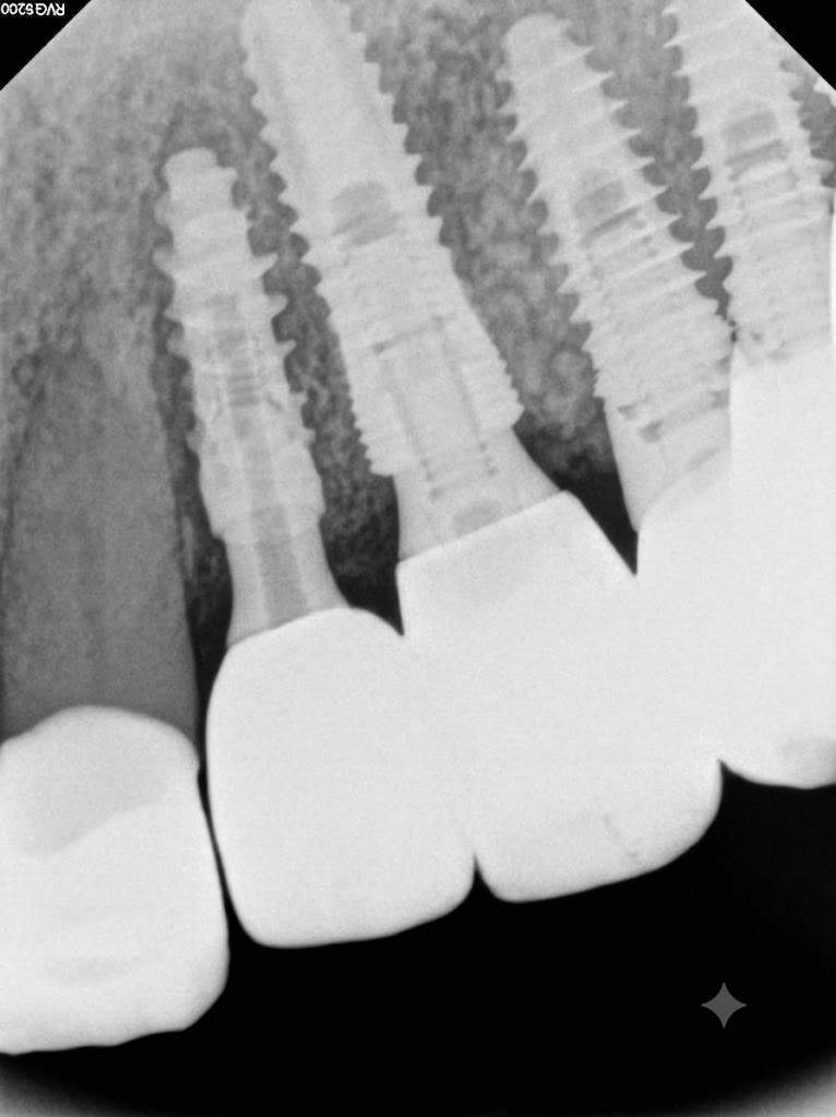
A single Ø2.5 × 10 mm drill was used according to the D032 bone quality, followed by radiographic verification of the implant position.


A Ø3.5 × 10 mm ALX-BT Narrow implant (3.2 mm neck diameter) was placed, achieving an insertion torque of 35 N/cm.


A healing abutment was connected.


Bone grafting was performed using bovine cancellous bone.


Implant stability was measured using AnyCheck, with an IST value of 76.

Post-operative intraoral photograph confirming optimal implant positioning in the maxillary anterior region.


3️⃣Prosthesis
An intraoral scan was performed 7 weeks after implant placement.


An intraoral scan was performed 7 weeks after implant placement.


Final panoramic and periapical radiograph


Comments