Immediate Placement of ALX-BT Implants in Mandibular Posterior Extraction Sockets


Case Summary 🔎

Patient Information
92y, Female
Medical history : Hypertension, Hyperlipidemia, Osteoporosis
Diagnosis
Treatment Plan
Immediate implant placement at both extraction sockets
Fixture placement: ALXBT 5008 at both sites
Bone grafting
Case Overview
This case involved immediate implant placement in the mandibular posterior region at #35 and #36 following same-day extraction. The teeth were indicated for extraction due to chronic periodontitis. After the initial drilling, bone density was assessed intra-operatively and identified as D333. A minimally invasive drilling protocol was performed using the ALX Kit. ALX-BT 5008 fixtures were placed at both sites, achieving initial torque values ranging from 70 to 40 Ncm and an IST value of 88. Bone grafting was performed using Neoss-B xenograft and autogenous bone to manage the defect areas. At 5 weeks postoperatively, final cementless zirconia prostheses were delivered, demonstrating an efficient treatment workflow from immediate placement to final restoration.
Case Presentation
1️⃣Pre-op
Teeth #35 and #36 presented with chronic periodontitis and were considered to have a hopeless prognosis, requiring extraction.
Immediate implant placement was planned based on the preservation of the buccal and lingual socket walls.

2️⃣ Surgery
Sequential drilling was performed from Ø2.5 × 10 mm to Ø4.5 × 10 mm.
Alveoloplasty was performed to recontour the sharp bony ridge.
Bone density was assessed after the initial drilling and identified as D333.
Osteotomy direction and depth were radiographically verified.
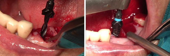
ALX-BT 5008 fixtures were placed at both extraction sites.
Initial torque was adjusted from 70 to 40 Ncm.

Healing abutments were connected to the placed fixtures.
Extraction socket defects were grafted using Neoss-B xenograft and autogenous bone. The graft material was adapted to support defect fill.

The flap was repositioned and sutured to achieve soft tissue closure around the healing abutments.
Postoperative periapical radiograph


3️⃣Prosthesis
Intraoral scanning was performed 3 weeks after surgery.
At 5 weeks postoperatively, final cementless zirconia prostheses were delivered with an IST value of 78 recorded before restoration.


Panoramic radiograph after final prosthetic delivery


Comments