S-Mini in the Lower Anterior Region for Implant Placement in a Narrow Ridge
- GAO
- Jun 16, 2025
- 2 min read

Updated: Jul 23, 2025
Case Summary 🔎
Patient Information
Age/Sex: 61-year-old Male
Systemic Condition: Hypertension (well-controlled with medication)
Clinical Findings
Tooth Site: #33
Oral Examination: Extensive apical pathology observed on #33
Diagnosis: Apical Periodontitis on #33
Clinical Summary
The patient presented with pain in the mandibular anterior region. Radiographic analysis revealed a severe apical lesion affecting tooth #33, indicating the necessity of extraction followed by implant placement. Given the extensive inflammation, a delayed protocol was chosen. An S-mini implant, optimized for anterior sites, was placed 7 months after extraction. Prosthetic restoration was completed with excellent functional and aesthetic outcomes. At the 8-year follow-up, the implant remained clinically and radiographically stable.
Treatment Plan
Extraction of tooth #33
Delayed implant placement
Final restoration using a Zirconia crown
Periodic follow-up evaluations
Conclusion
Placement of S-mini implant in the mandibular left canine
Final restoration with Zirconia crown
Periodic follow up with stable long term outcomes
Case Presentation
1️⃣ Pre-Op
Radiographic examination revealed a severe apical lesion on tooth #33, indicating the need for extraction and subsequent implant placement.

2️⃣ Extraction & Healing
After extraction, inflammatory tissue was thoroughly debrided. Bone grafting was performed using Regenoss (allograft), and the site was covered with a pedicle flap to support soft tissue healing.


TissueBond was applied to stabilize the flap and assist in maintaining soft tissue volume and supporting gingival contours.


At 5 months post-extraction, soft tissue healing and partial resolution of inflammation were observed clinically and radiographically.


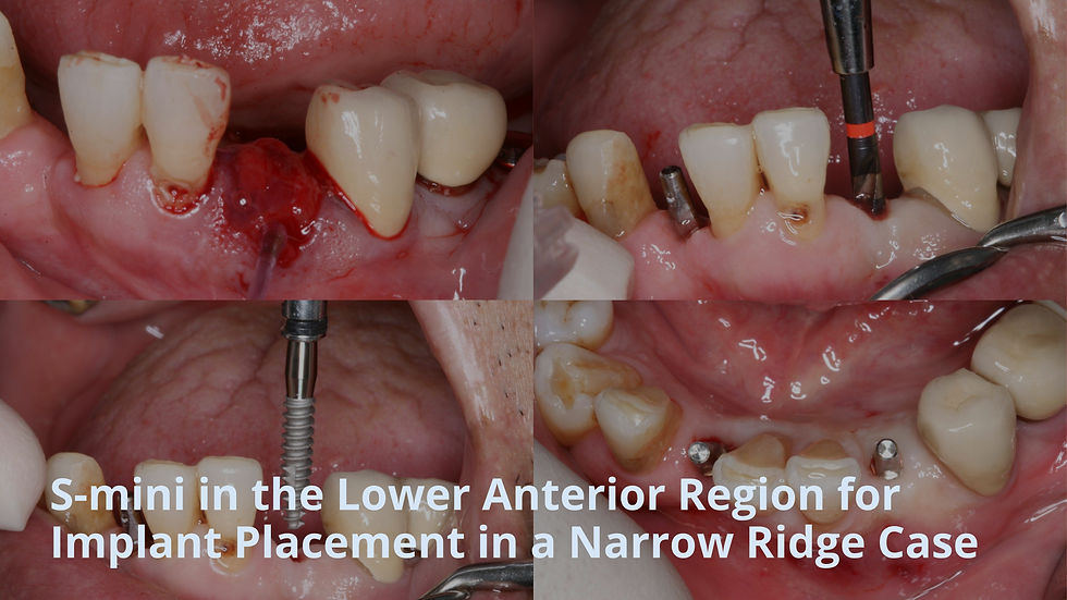
3️⃣ Implant Placement in a Narrow Ridge
7 months post extraction, good healing and healthy gingival contours were observed.
Using the S-mini kit and drills, depth-controlled drilling was performed based on the reference markings in a flapless approach.


∅3.0*13mm S-mini implant was placed.

The implant was accurately placed in the ideal position without exposure, despite the narrow ridge width.


S-mini Fixture
S-mini is a one-body implant designed for narrow ridges, such as the mandibular anterior region. It is available in diameters of 2.5, 3.0, and 3.5 mm, and is placed using a dedicated surgical kit for precise and safe placement.

4️⃣ Post-Op
Post-operative radiographs shows well positioned implant.


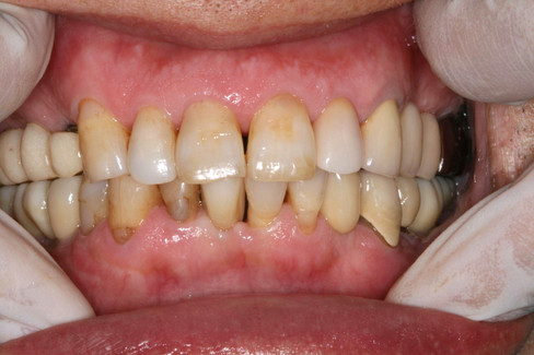
5️⃣ Final Restoration
The final prosthetic restoration was delivered 5 months after surgery.


6️⃣ Follow-Up
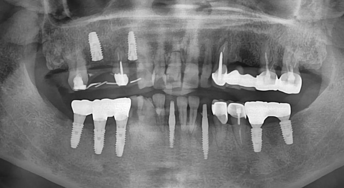
The 1-year follow-up panoramic radiograph and intraoral photograph show good maintenance with no signs of complications


The 3-year follow-up panoramic radiograph and intraoral photograph.


At the 8-year follow-up, clinical and radiographic evaluations revealed well-maintained peri-implant tissues with no signs of marginal bone loss.


Comments