Delayed re-implantation using ALX and Prosthetic restoration with CL-Link
- GAO
- Nov 7, 2025
- 2 min read

Case Summary 🔎
Patient Information
73 y/o, Male
Past Medical History: Healthy
Introduction
This case report describes the delayed re-implantation of a mandibular posterior site using the ALX implant system, followed by digital prosthetic restoration with the CL-Link system. The patient presented with peri-implant bone loss around the distal implant of a mandibular implant-supported bridge. The compromised implant was removed, and bone grafting was performed. After an appropriate healing period of approximately four months, a new implant was placed using the ALX system. About four weeks later, a digital impression was captured using an intraoral scanner, and the final prosthesis was delivered with the CL-Link system. At the follow-up three months after prosthesis delivery, the restoration remained stable without complications. This case highlights a structured approach to managing peri-implant complications through delayed re-implantation and a fully digital workflow using ALX and CL-Link.
Treatment Plan
Removal of the existing #35–37 implant-supported bridge and crown sectioning at #37
Fixture removal at site #37 and bone grafting for ridge preservation
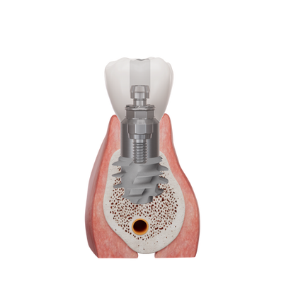
After 4 months of healing, implant placement at #37 using the ALX system
Digital impression taking 2 weeks after implant placement
Final prosthetic restoration using the CL-Link
3-month follow-up
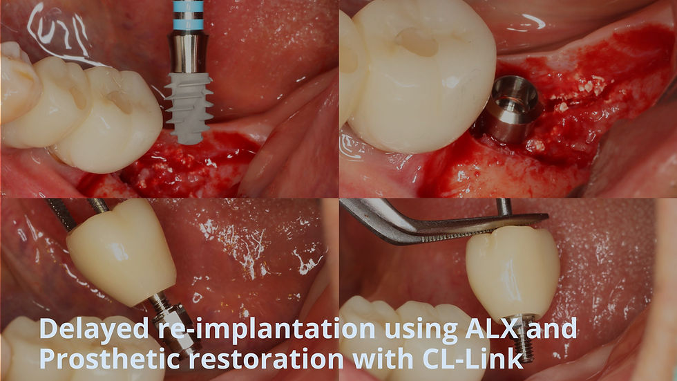
Case Presentation
1️⃣Pre-op
Pre-operative radiograph shows peri-implant bone loss around the #37


#35-37 implant bridge was removed.


2️⃣ Surgery
#37 implant was removed and bone graft was placed for ridge preservation.


Grafted ridge observed after fixture removal and bone grafting.


Well-preserved ridge with adequate bone volume observed 4 months after grafting.


With drilling path confirmed, sequential drilling was completed and Maxy was performed.


ALX-IT 5.5x8mm, cuff 4mm implant was placed at 40N/cm, achieving an 73 ISTV.


Bone grafting with allogeneic bone was performed and covered with a resorbable collagen membrane, followed by suturing.


Post operative radiograph


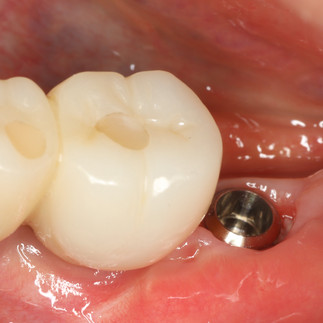
IST value remained stable at 2-week follow-up, and digital impression was taken using an intraoral scanner.


3️⃣Prosthetic Fabrication
IST value remained stable at 2-week follow-up, and digital impression was taken using an intraoral scanner.


4️⃣ Final restoration
Final prosthesis fabricated with the CL-Link was delivered 4 weeks after impression, 6 weeks post-surgery.


Intraoral photograph and periapical radiograph show the final prosthesis seated with proper fit and stability using the CL-Link.


5️⃣Follow Up
At 3 months, the prosthesis remains functional and peri-implant bone levels are well-maintained.


Comments