Flapless Early Loading of Short and Wide ALX Implants in the Maxillary Sinus Region
- GAO
- Oct 14, 2025
- 2 min read


Case Summary 🔎
Patient Information
Age/Sex: 73-year-old / Female
Past Medical History: History of Osteoporosis
Introduction
The patient presented with inflammation in the #27 region, necessitating removal of the existing bridge and extraction of tooth #27 due to chronic apical pathology. Residual bone height at site #26 was approximately 4-5 mm, and a sinus lift was performed using the SCA (Sinus Crestal Approach) technique without grafting, followed by placement of an ALX implant. Five days post-placement, a digital impression was taken, and the definitive prosthesis was delivered two weeks after surgery, following an early loading protocol.
Short and wide ALX implants were used in the maxillary posterior region, including a sinus-lifted site without grafting. Despite limited residual bone, final prosthetic loading was successfully completed within two weeks, demonstrating clinical stability and the predictability of this minimally invasive approach.
Treatment Plan
1) Extraction of #26
2) Implant placement at #26,27
3) Digital impression
4) Final restoration with Zirconia crown
5) 1 month follow up at #26,27
Case Presentation
1️⃣ Pre-Op


2️⃣ Surgery
A VAROGuide was fabricated and used for guided implant surgery. To facilitate a flapless procedure, soft tissue was removed at the implant site using a tissue punch through the surgical guide.
A sinus drill from the VAROGuide Sinus Kit was used with a 3 mm stopper for initial osteotomy preparation.
The S-reamer from the VAROGuide Sinus Kit was used to drill the remaining bone until the cortical bone of the sinus floor was reached.


The residual bone height was measured using a depth gauge to confirm the available bone.
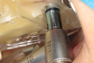
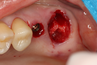
Ø6.0 × 6 mm, Cuff 2 mm ALX-IT, short and wide implant was placed at site #26 with D442 bone density.


An implant was placed following sinus lifting without grafting. (Class II sinus classification)


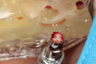
Healing abutments were connected for ISTV measurement, and initial stability was assessed using the AnyCheck device. (#26 : 81+2, #27 77+2)

Bone grafting was performed at site #27 using xenograft material.(Neo oss-B, S1 bone)


3️⃣ Post-Op
A digital impression was taken using an intraoral scanner.


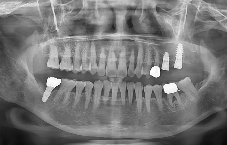
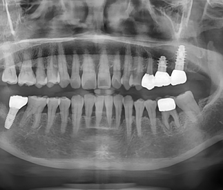
Post-operative panoramic radiograph
4️⃣ Final Restoration
Two weeks post-surgery, soft tissue healing was observed, and the IST value remained stable at 80.


The final prosthesis was delivered using the CL Link.


The screw access hole of the crown was sealed with resin
Intraoral photograph and periapical radiograph after final restoration


1 month follow-up showing stable soft tissue and well-maintained implant restorations.


Comments