Re-implantation of Failing Implants Using ALX System and Final Restoration with CL-Link
- GAO
- Oct 28, 2025
- 3 min read


Case Summary 🔎
Patient Information
77 y/o, Female
Past Medical History: Osteoporosis (taking oral medication once a week)
Dental History: Multiple implants previously placed on both arches; recurrent peri-implantitis and bone loss noted around maxillary left posterior implants
C.C.: “I feel discomfort and swelling around the upper left implants”
Diagnosis: Peri-implantitis with severe bone defect in the maxillary left posterior area (#25–27)
Introduction
A 77-year-old female patient with a history of osteoporosis presented with discomfort and swelling in the maxillary left posterior region. Clinical and radiographic examination revealed advanced peri-implantitis around previously placed implants, accompanied by significant bone loss and soft tissue inflammation.
Considering the compromised bone quality due to both peri-implantitis and systemic bone metabolism, a comprehensive treatment plan was established focusing on complete removal of the failing implants, guided bone regeneration, and re-implantation using the ALX system known for its excellent initial stability in poor bone conditions.
After a sufficient healing period, final restoration was completed using the CL-Link system, providing a precise and stable prosthetic connection while ensuring functional recovery and esthetic harmony.
Treatment Plan
Removal of existing abutments and failed fixtures (#25–27)
Thorough debridement and bone grafting for ridge reconstruction
Re-implantation after 5 months using ALX fixtures (#25: ALXIT44510, #26: ALXIT55010, #27: ALXIT55010)
Placement of healing abutments and soft tissue maturation
Intraoral scanning and fabrication of final prosthesis using CL-Link system
Delivery of final prosthesis and post-restorative evaluations
Case Presentation
1️⃣ Pre-Op
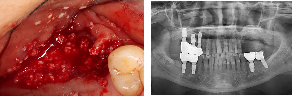
Intraoral photograph and panoramic radiograph taken on the first visit showing severe peri-implant bone loss around the #25-27, with the fixture threads exposed. Replacement with new fixtures was planned after removal of the existing failing fixtures

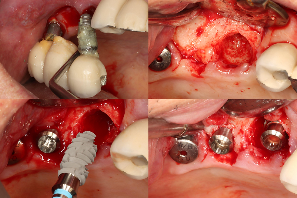
Using a bur to section and remove the existing prosthesis around #25-27 in preparation for fixture removal
After bridge removal, the remaining abutments on #25-27 are exposed

Bone graft materials was placed to restore the alveolar ridge and promote regeneration before re-implantation
The significant bone defect is clearly visible in the panoramic radiograph
2️⃣ Surgery
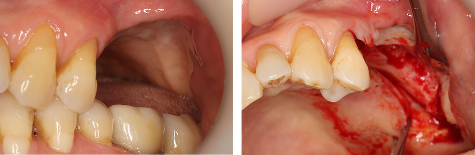
5 months after grafting, the edentulous area shows stable soft tissue healing with adequate keratinized gingiva
A full-thickness flap was elevated to expose the alveolar ridge at the #25-27 area. The grafted bone appears well-maintained with sufficient ridge width and height
An ALX-IT 4.5 x 10mm, cuff 4mm was placed at #25, with D221 bone density
An ALX-IT 5.0 x 10mm, cuff 5mm was placed at #26, with D332 bone density

ALX-IT fixtures were placed. #25 - ALX-IT 4.5 x 10mm, cuff 4mm, D221, 40N/cm
#26 - ALX-IT 5.0 x 10mm, cuff 5mm, D332, 30N/cm
#27 - ALX-IT 5.0 x 10mm, cuff 5mm , D332, 30N/cm
After implant placement, bone grafting was performed around the exposed threads and between the fixtures to enhance bone volume and achieve stable ridge contour

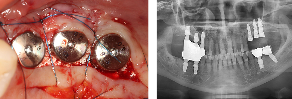
After placement of all 3 fixtures and bone graft material, suture was done
Post-op panoramic radiograph showing the placement of 3 ALX fixtures with ideal positioning and parallelism
3 months post-surgery, the healing abutments were removed, revealing well-healed soft tissue with healthy contours around the implant site
Scan posts were connected to the 3 fixtures to capture accurate digital impression

Scanning view
The soft tissue around the implants showed excellent healing and stable gingival contour, providing a healthy foundation for the final restoration

3️⃣ Final Restoration
The final prosthesis was fabricated using the CL-Link system, allowing for a precise and passive fit on the 3 ALX fixtures.
Buccal view after final prosthesis delivery shows harmonizing with adjacent natural teeth

Occlusal view shows excellent crown morphology and harmonious occlusal contact
Final panoramic radiograph after prosthesis delivery shows 3 ALX implants (#25-27) with well-integrated bone support and accurately seated restoration

Conclusion
This case demonstrates the successful management of peri-implantitis and bone defect in a 77-year-old osteoporotic patient through the use of ALX implants and final restoration with CL-Link.
After removal of the failed fixtures and thorough bone grafting, the re-implanted ALX fixtures achieved excellent primary stability even in compromised bone conditions. The subsequent healing period allowed for sufficient osseointegration and soft tissue maturation, resulting in a favorable surgical and prosthetic outcome.
The final prosthesis fabricated with the CL-Link system provided accurate fit, functional stability, and esthetic harmony, restoring the patient’s masticatory function and comfort.
This case highlights the clinical reliability of ALX implants in re-implantation cases and the precision of the CL-Link system in achieving a long-term, predictable restorative result, even in elderly patients with systemic bone metabolism concerns.


Comments