Full-Arch Rehabilitation using ALX-IT implants
- GAO
- Sep 30, 2025
- 3 min read

Updated: Oct 2, 2025

Case Summary 🔎
Patient Information
56 y/o, Female
Past Medical History: No systemic disease reported
Dental History: Remaining teeth with poor prognosis and several failed implants present
C.C.: “I cannot chew due to multiple missing and failing teeth”
Diagnosis: Edentulous condition planned after extraction all remaining teeth and failed implants
Introduction
A 56-year-old female patient presented with a chief complaint of being unable to chew properly due to multiple missing and failing teeth. Clinical and radiographic examination revealed remaining dentition with poor prognosis as well as previously placed implants showing signs of failure. Considering the patient’s desire to regain full masticatory function and improve quality of life, a treatment plan was established to perform full-arch rehabilitation.
All remaining teeth and failed implants were scheduled for removal, followed by placement of ALX implants to restore function and esthetics. The case highlights the use of ALX implants in managing an edentulous condition, providing immediate stability and long-term predictability for full-arch restoration.
Treatment Plan
Extraction of remaining teeth and removal of failed fixtures
Placement of ALX implants in both maxilla (6 fixtures) and mandible (4 fixtures) for full-arch rehabilitation
Immediate loading with a full-arch implant-supported fixed temporary prosthesis
Delivering of final prosthesis
Case Presentation
1️⃣ Pre-Op
Panoramic radiograph and intraoral view showing multiple remaining teeth with severe caries, failing prostheses, and exposed implant fixtures

2️⃣ Surgery (Maxillary arch)
Initial stage of extraction and removal of previously placed implant fixtures, showing remaining teeth with severe decay
Surgical site after extraction and removal of failed fixtures, showing exposed alveolar bone and preparation for implant placement

Subsequent drilling performed under irrigation for ALX implant site preparation
Osteotomy performed with MAXY drill to create the final shape matching the ALX thread design before fixture placement

AN ALX-IT 4.5 x 12mm, cuff 3mm was placed at #11, achieving 40N/cm of insertion torque
ALX fixtures placed with healing abutments connected, and bone graft applied to deficient ridge areas

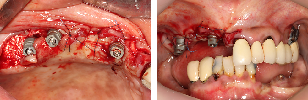
Scan bodies connected to MUA abutments with sutures completed after ALX implant placement and bone grafting
Intraoral view after ALX placement with MUA scan bodies in the left maxilla, while existing fixture and prosthesis remain in the 2nd quadrant prior to removal
Removed prosthesis and failed teeth from the 2nd quadrant, showing severe caries and restoration failure
Surgical site after extraction and fixture removal, showing irregular alveolar ridge and unfavorable bone level

Subsequent drilling was performed under irrigation like 1st quadrant, then drilling with MAXY to create the final shape matching the ALX thread design
ALX-IT fixtures were placed. Dehiscence defects are observed with exposed threads. #21: ALX-IT 4.5 x 12mm, cuff 3mm
#24: ALX-IT 4.5 x 12mm, cuff 3mm
#26: ALX-IT 5.5 x 10mm, cuff 3mm

3️⃣ Surgery (Mandibular arch)
Intraoral view of the mandibular anterior region before extraction, showing remaining teeth with severe periodontal involvement then extraction of remaining teeth
Drilling with MAXY after using twist surgical drill then placement of ALX-IT fixtures

ALX-IT were placed in the mandible, showing stable distribution across the anterior and posterior
#46: ALX-IT 4.5 x 8mm, cuff 3mm
#42: ALX-IT 4.5 x 8mm, cuff 3mm
#31: ALX-IT 4.5 x 8mm, cuff 3mm
#36: ALX-IT 4.5 x 8mm, cuff 4mm
Post-op panoramic radiograph

4️⃣ Prosthetic Part
Intraoral view at 5 days post-surgery, prior to placement of the full-arch implant-supported fixed temporary bridge in the maxilla
Final view showing both maxillary and mandibular implant-supported fixed temporary bridges in place, restoring esthetic and function

After 4 months, scan bars were connected for accurate intraoral scanning
Scanning view

5️⃣ Final Restoration
Intraoral view and panoramic radiograph showing completed full-arch final restorations in both maxilla and mandible, achieving functional and esthetic rehabilitation

Conclusion
This case demonstrates the successful full-arch rehabilitation of an edentulous patient using ALX implants. By removing all remaining teeth and failed implants, and strategically placing six maxillary and four mandibular ALX fixtures, stable function and esthetics were restored.
The use of ALX implants provided excellent primary stability and allowed for immediate loading with a temporary prosthesis, ensuring that the patient could regain masticatory function and confidence in daily life without delay. After healing, the delivery of definitive full-arch prostheses achieved both long-term predictability and improved quality of life.
This case highlights the clinical effectiveness of ALX implants as a reliable solution for managing complex edentulous conditions with failing dentition and previous implant failures.


Comments