Final Crown in Two Days after ALX Implant Placement in Sloped Alveolar Ridge
- GAO
- Jul 23, 2025
- 2 min read

Updated: Jan 23
Case Summary 🔎
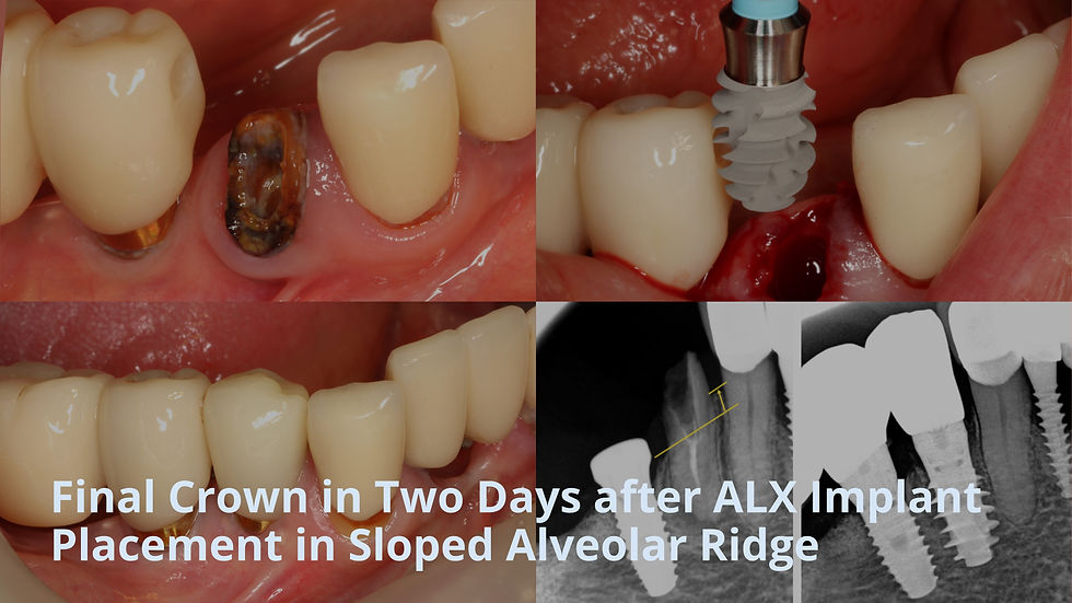
This case involved managing a 2 mm oblique alveolar ridge at site #44, where the mesial bone was higher than the distal. A tissue‑level ALX implant was used to navigate this anatomy without flap or graft procedures, enabling precise platform placement.
The ALX Implant’s narrow-core, deep-thread self‑compaction design facilitated stable insertion in the sloped ridge, achieving high primary stability (ITV 40 Ncm, ISTV 80). The availability of a 3 mm cuff, fitting the deeper distal bone, helped create a smooth soft tissue emergence without overexposing the implant platform.
Delivery of the final zirconia crown after two days resulted in a stable occlusion, and five-month follow-up confirmed healthy soft tissue and bone preservation.


Case Presentation
1️⃣ Pre-Op
The patient presented with a crown fracture at site #44, extending subgingivally with insufficient structure for restoration, making extraction necessary.
The treatment plan included extraction, immediate implant placement, and definitive crown delivery within 48 hours.


A preoperative panoramic radiograph showed dentinal caries in the residual root, confirming the tooth was unrestorable.

The alveolar ridge had a 2 mm height difference between mesial and distal sides, requiring consideration for implant depth and platform positioning.

2️⃣ Surgical phase - ALX Implant Placement
Tooth #44 was atraumatically extracted to preserve surrounding bone and soft tissue.


Site #44 was prepared using a stepwise drilling sequence, from initial to final drill, considering bone density D222 (D2L2LB2LB2).


An ALX IT35508WT implant was placed at site #44, with insertion torque adjusted from 70 to 40 Ncm, and ISTV recorded at 80.
Considering the 2 mm vertical bone discrepancy between mesial and distal, the implant platform was aligned with the distal bone crest, and a 3 mm cuff was selected for optimal soft tissue emergence.


A YK abutment was connected to the implant following placement.


A scan body was connected on the YK abutment to facilitate digital impression taking.

A postoperative panoramic radiograph was taken to confirm implant position and angulation.

3️⃣ Prosthetic phase
The final prosthesis was delivered two days after surgery, consisting of a zirconia crown fabricated on a YK abutment.


Final occlusal adjustment was performed to ensure stable and balanced contact.


A panoramic radiograph taken after crown delivery confirmed proper marginal fit and overall implant alignment.

CBCT was also performed to assess the three-dimensional position and bone adaptation around the implant.

Video
4️⃣ Follow-Up
At five months post-op, the implant remained stable with healthy surrounding soft tissue and no signs of inflammation or bone loss.


Comments