Early placement of the Neobiotech IS II Implants on the maxillary posterior area
- Dr. Yong Soo Kim

- Nov 2, 2015
- 1 min read
Situation
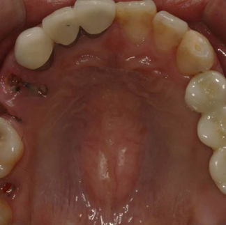
26 year old male patient presented with multiple carious lesions, multiple amalgam fillings, and PFM crowns on #11 & 21.
Pre-operative Observation


Treatment Plan
1. Scaling and root planing
2. Extraction of the tooth #24, 25, 26, 27, 37 and 38
3. Root canal therapy and Crown fabrication of the tooth #36
4. Implant placement of #24,25 and 26 at 2 month after extraction with simultaneous guided bone regener5. ation
6. Uncovering at 4 months after the implant placement
7. Final prosthesis

2 months after extraction of the tooth #24, 25, 26 and 27.

Extraction site was not completely healed.

Implant direction pin was placed.

Neobiotech SLA Implants were placed and primary stability was achieved.

FDBA was grafted on the defect of the site.

Grafted site was covered by collagen membrane.

Flaps were sutured by using both horizontal mattress suture and single interrupted suture.

Implant placement of the tooth #24,25,26 with simultaneous guided bone regeneration – panorama

4 month post-op

After 4 months, incision was made for the uncovering.

Both buccal and palatal full thickness flap were raised.

Complete healing of the extraction site was noted.

Healing abutments were placed.

Uncovering after 4 month

Pick-up impression was taken.

Abutments were placed and tightened to 35Ncm.

3- unit splinted SCRP implant zirconia crown has been fabricated.
Post-operative Intraoral photograph

Neobitech SLA implant has shown to be successfully placed on the post extraction site with a good primary stability.


Comments