Sinus Class III CM and Class IV CM/C Fixations with SCA and SLA Techniques
- Dr. Jongyub Kim

- May 31, 2018
- 4 min read
Patient information A 54 year-old male, well-controlled hypertension and was taking a beta-blocker prescribed by his family doctor.
Preoperative observation
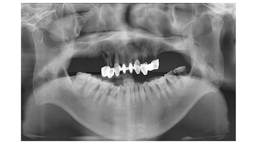
Panoramic radiograph at the time of first visit showing severe periodontitis and vertical bone loss at teeth #12, 13, 14 and 22, and limited residual bone on both sides.

Clinical view of frontal side at first visit (2008-10-16).
Maxillary front FPD was mobile. and severe anterior deep bite was seen. Also pus discharge from gingival sulcus of #9 was found.


A CT radiograph of the right maxillary posterior area showing 3-5mm height of residual bone and healthy sinus cavity. A CT radiograph of left maxillary posterior area showing 5-6mm height of residual bone and healthy sinus cavity.

Frontal view in occlusion two weeks after extraction of #12,13,14, and 22. Severe vertical bone loss at the extraction sites and severe extrusion of lower anterior teeth were observed.

Frontal occlusal view. Broad posterior alveolar bone tables, and severe bone deficiency in the right canine area were observed.

Buccal view of right side at the first visit.

Buccal view of left side at the first visit. Broad alveolar ridge was observed.

Occlusal view of right maxillary posterior area. Broad crestal alveolar ridge and adequate attached gingiva were observed at #14-17 missing area, but severe deficiency seen at #13 area. Denture soreness was seen at posterior buccal area.

Occlusal view of left maxillary posterior area. Broad crestal alveolar ridge and adequate attached gingiva were observed at #26 and 27 missing area.

Enlargement osteotomy made up to 3.8mm in diameter with the 4mm stopper as the residual bone height was estimated to be at least 5mm.

3.2mm S-reamer was used to open the inferior cortical wall of the sinus starting with the 5mm length of stopper. The drilling depth was increased 1mm by 1mm until the reamer met the inferior border of the maxillary sinus. The diameter of the S-reamer was chosen based on the diameter of an implant engaged at the sinus inferior wall to obtain an ideal insertion torque and fixation.

The narrower flat end depth gauge in the SCA kit was utilized in order to check the opening of the inferior bony wall of the maxillary sinus. The depth gauge should be inserted with tactile sensation by touching the lateral wall of the osteotomy.
When the depth gauge reaches the floor and if the floor is blocked (not opened) by bone, keep drilling 1mm more and check again until the floor wall is opened. Height of residual bone was measured with the depth gauge by hanging on the inferior wall.

Before inserting bone graft materials, membrane tearing was checked by asking the patient to blow air through nose (Valsalva Maneuver). Bone grafting could be conducted if membrane was not torn. Bone graft material (Calpore 0.5cc) was inserted into the osteotomy to the maxillary sinus using bone inserter and spreader. It is recommended to stop inserting bone graft materials when certain pressure from grafting materials in the maxillary sinus can be felt, which can represent the tension of the maxillary sinus membrane.

Panoramic radiograph showing 2 implants placed after bone grafting at #26, and 27 area. About 5mm sinus lift was performed and graft materials are observed as dome-shaped radiopaque mass around periapical area of implant at #27.
Fixation types were Class II C 3 M 3 I 2 fixation with 40 Ncm at #26 and Class III C 3 M 2 fixation with 30 Ncm.

Right side of the maxilla at #16 and 17 missing area was grafted through the SLA technique. Small bluish colored half-rounded area seen just above the maxillary sinus membrane. Bony window was made first by one side of the LS-reamer. The membrane could be detached through the small opening before making an entire round hole with the reamer.

3 implants (CMI IS fixtures) were placed by flapless surgery after sinus bone graft at #15, 16, and 17 area.

Fixation types were D333 with 30 Ncm, D320 with 30 Ncm, and D200 with 25 Ncm for the #15, 16, and 17 area respectively.

Panorama radiograph showing right and left maxillary posterior area after the surgery. Bone graft materials were observed as dome-shaped radiopaque mass around implants of #16, 17 and 27.

CT scan at #16 and 17 after the surgery showing graft materials well positioned as dome shape around the implants. If implants were placed simultaneously, lifting up grafting materials more than the length of the implant would not be needed.

Occlusal view at 4 months after the surgery. Sufficient attached gingiva was observed.

Clinical photograph at 4 months after the surgery. Soft tissue was well healed.

Panoramic radiograph at 8 months after the implant placement. #17-16-13-22 and 26-27 implants had been loaded with provisional restorations.

Panoramic radiograph at 13 months after implant placement. Surrounding bone and graft materials around the implants were well maintained.

Buccal view of maxillary right posterior area at the time of final prosthesis delivery.

Buccal view of maxillary left posterior area at the time of final prosthesis delivery.

Periapical radiographs of right and left maxillary posterior area at 13 months after implant placement. Marginal bone around implants was well maintained.

Periapical radiographs of right and left maxillary posterior area at 13 months after implant placement. Marginal bone around implants was well maintained.

Panoramic radiograph at 7 year follow-up. Surrounding bone and grafting materials around implants are being well maintained as before.

Periapical radiograph at 8 years after implant surgery. Marginal bone around implants had been well maintained and has even over-grown.

Periapical radiograph at 8 years after implant surgery. Marginal bone around implants had been well maintained and has even over-grown.

Clinical photograph of right posterior maxilla at 8 year follow-up check.

Clinical photograph of left posterior maxilla at 8 year follow-up.

8 year follow-up CT scan at #16, 17, 26 and 27 implants showing bone around the implants had been well maintained.


Comments